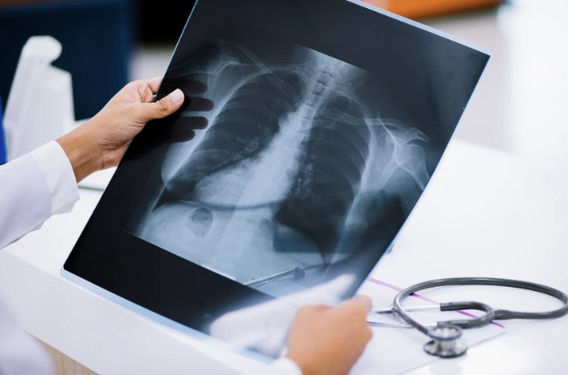
Lung cancer is one of the deadliest forms of cancer in America, with 125,000 deaths estimated to come in 2025 alone.
However, patients diagnosed in earlier stages have a significantly higher likelihood of better outcomes. In response, doctors across the nation are constantly searching for new treatment methods to help lung cancer patients get on the road to recovery.
Luckily, a recent innovation in treatment technology has opened the door for a new type of procedure – one that combines diagnosis and nodule removal into one visit as a single anesthesia event.
At the forefront of this cutting-edge initiative is Capital Health Cancer Center, whose multidisciplinary team is currently one of the only groups in the region offering this type of surgery. We’ll cover exactly how that works below.
The Latest Treatment: Diagnosis and Removal of Lung Cancer Nodules During a Single Surgery
Traditionally, when a lung nodule is discovered, the patient is referred to a pulmonologist who will need to run two to three separate procedures as part of their assessment:
- A biopsy to extract tissue for sample analysis
- A second biopsy on the chest’s lymph nodes to determine if the cancer has metastasized
- A surgery to remove the lung nodule if it’s deemed cancerous
With each of these procedures done separately, patients must deal with the stress and anxiety that comes with waiting – a feeling that’s only exacerbated by the frustration of being back under the scope a second and third time if necessary. For some, this wait can take up to three months from discovery to treatment.
However, through the revolutionary use of robotic, minimally invasive technologies, each of these procedures can now be combined into one single anesthesia event.
What Is the Procedure Like?
The procedure involves a multidisciplinary approach, tapping into cancer experts of all kinds who are staffed here at Capital Health: an interventional pulmonologist, anesthesiologist, pathologist, and thoracic surgeon.
Once the patient is under general anesthesia, their doctor can perform a minimally invasive biopsy of the nodule and the lymph glands in their chest using state-of-the-art technology. This technology allows the team to determine whether a nodule is cancerous and/or whether the cancer has appeared to metastasize in the lymph nodes.
If the nodule is deemed benign, the procedure ends, helping the patient avoid unnecessary follow-up surgeries.
If the nodule is deemed malignant, the surgical team can immediately and precisely remove it through robotic-assisted surgery.
Afterward, the patient is woken up and briefed on how the procedure went. The doctor will work together with the patient to help determine the next steps and establish a plan for the future if any additional treatment is required.
Robotic-Assisted Surgeries
Da Vinci Robotic–Assisted Surgical System
Operated through a specialized robotic console, the da Vinci® Assisted Surgical System allows trained surgeons to guide ultra-precise surgical instruments with movements that are smoothly translated by the machine in real time.
Its high-definition camera provides magnification of up to 10 times what the human eye can see, delivering an unparalleled view of the surgical area and allowing surgeons to see even the smallest details with exceptional clarity. Additionally, the system’s enhanced range of motion mimics the dexterity of the human hand but far exceeds its natural capabilities, facilitating complex actions such as suturing, knot-tying, and tissue removal with remarkable efficiency.
The precision of the da Vinci system not only reduces the impact on surrounding healthy tissue but also promotes quicker recovery times for patients. By incorporating this cutting-edge technology, Capital Health ensures that patients benefit from safer procedures, improved outcomes, and a level of care that exemplifies its commitment to quality and innovation.
Ion Robotic Bronchoscopy
The Ion Robotic Bronchoscopy system represents a significant leap forward in the diagnosis and management of lung conditions – particularly lung cancer.
Equipped with an ultra-thin, highly maneuverable catheter, the Ion system allows doctors to access all 18 segments of the lungs with unprecedented precision, even reaching nodules located in the most hard-to-reach peripheral regions. By utilizing advanced shape-sensing technology, this system ensures real-time accuracy in navigation and biopsy tool placement, effectively improving diagnostic accuracy while minimizing risks. Patients benefit greatly from the minimally invasive nature of this procedure, which reduces discomfort and promotes faster recovery times for the patient.
At Capital Health, the adoption of the Ion Robotic Bronchoscopy not only enhances our ability to diagnose lung conditions at earlier stages but also streamlines the diagnostic process, enabling patients to receive timely, effective care. This advanced technology empowers our expert medical team to deliver a higher standard of care, ultimately improving patients’ quality of life with faster diagnoses and better treatment outcomes.
Trust the Team at Capital Health
If you or someone you know thinks they may be showing symptoms of lung cancer, do not wait to take action – earlier stages of cancer are much easier to treat, and it’s always better to err on the side of caution.
To schedule a screening appointment, simply visit our website or give our offices a call at 609-537-6363. Our multidisciplinary team will work together with you to make sure you get the best care available, no matter your situation.
Capital Health Cancer Center, located in Hopewell, New Jersey, is home to one of the nation’s Lung Centers of Excellence as well as other centers of excellence specializing in breast care, liver health, neuro-oncology, pancreatic health, and robotic-assisted surgery. To learn more, visit capitalhealthcancer.org.